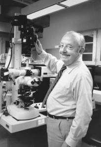
 The Keith R. Porter Imaging Facility is named for the former chair of the Biological Sciences Department (1984-1988), who was an eminent electron microscopist.
From the Journal of Cell Biology memorial article:
“Keith Roberts Porter died on May 2, 1997, just over a month short of his 85th birthday. He had the perspicacity, good fortune, and patience to take advantage of the fast moving frontier of analytical biology after the Second World War to provide many of the techniques and experimental approaches that established the new field of biomedical research now known as cell biology. He was renowned for taking the first electron micrograph of an intact cell, but his contributions went far beyond that seminal instance. They ranged from technical developments, such as the roller flask for cell culture and the Porter-Blum ultramicrotome, to experimental and observational achievements, such as studies on the synthesis and assembly of collagen, on the role of coated vesicles in endocytosis, on lipid digestion in the intestine, and on the universality of the 9 + 2 axoneme in cilia. The initial ultrastructure descriptions of the endoplasmic reticulum and the sarcoplasmic reticulum, identification of the role of T-tubules in excitation–contraction coupling in muscle and the role of the cytoskeleton in cell transformation and shape change, were his, as were many other contributions, described in some detail elsewhere (Peachey and Brinkley, 1983; Moberg, 1996). Absent from this list are his early pioneering work establishing the androgenetic haploid in frogs, an exercise in nuclear transplantation with consequences for the recent cloning of mammals, and his later adventures with pigment migration in fish chromatophores.”
For a complete biography, please refer to Wikipedia.